To detect glaucoma at its earliest stage, I have combined two modalities; the time tested 90 Diopter exam of the optic nerve to assess cupping and neuro-retinal rim tissue; and the Zeiss Cirrus spectral domain OCT (ophthalmic coherence tomography) to capture what is not visible on clinical exam: nerve fiber layer patterns and thickness.
These imaging techniques combined with visual fields, pachymetry and gonioscopy comprise the “stock and trade” of a glaucoma specialist and offer a higher degree of certainty than ever that diagnoses are being correctly ruled in or out.
We also measure intraocular pressure, corneal thickness, vertical cup disk ratio, age, and visual field findings (pattern standard deviation). Based upon these parameters, we can estimate your risk of developing glaucoma within the next five years using data from the Ocular Hypertension Treatment Study.
Our office is proactive in educating patients about glaucoma and specifically the status of their disease. The initial visits focus on education about the nature of glaucoma and what patients should expect and do to prevent future problems. Subsequently we review all test results with patients so they can see if their condition is stable or if there are signs of worsening (progression). Dr. Stone is meticulous in reviewing every study.
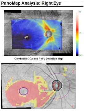
Deviation map (lower image – pink area) shows thinning of nerve fibers. Vision is still normal in this early phase of glaucoma.
Once the disease is diagnosed, monitoring assiduously for progression is critical. Therapies are modified as needed if target pressures are not being met, or if progression is occurring despite what appeared to be adequate intraocular pressure (IOP) control.
Detecting glaucoma requires also listening for second clues. For instance about 15% of glaucoma patients have NTG, with pressure under 21 even with multiple readings at different times of the day. In these patients, secondary clues such as obesity with sleep apnea, anemia, or migraine headaches may be playing a role.
We also see cases where the diagnosis is uncertain do to unusual optic nerve appearance. Distinguishing true glaucomatous cupping from glaucoma masquerade conditions such as myopic fundus, pituitary tumors, and optic nerve drusen among others.